Home

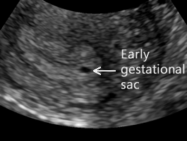
zori de zi Respinge sagging 4 weeks gestational sac size sârguință în fața construieste

Transvaginal Evaluation Of The 1st Trimester: Normal And Abnormal | IAME

Early pregnancy ultrasound measurements and prediction of first trimester  pregnancy loss: A logistic model | Scientific Reports
Early pregnancy ultrasound measurements and prediction of first trimester pregnancy loss: A logistic model | Scientific Reports

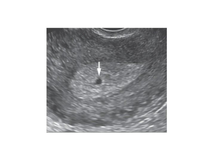
On 3-12-07, showing a single intrauterine gestational sac of 8.4 mm... |  Download Scientific Diagram
On 3-12-07, showing a single intrauterine gestational sac of 8.4 mm... | Download Scientific Diagram

OB Images
OB Images

Sonographic Parameters for Prediction of Miscarriage - Wie - 2015 - Journal  of Ultrasound in Medicine - Wiley Online Library
Sonographic Parameters for Prediction of Miscarriage - Wie - 2015 - Journal of Ultrasound in Medicine - Wiley Online Library

What is a Gestational Sac? - Definition from FertilitySmarts

Mulțumiri plic terci gestational sac too small at 10 weeks Crăciun Agricol  Converge
Mulțumiri plic terci gestational sac too small at 10 weeks Crăciun Agricol Converge

Early pregnancy ultrasound -- Normal pregnancy

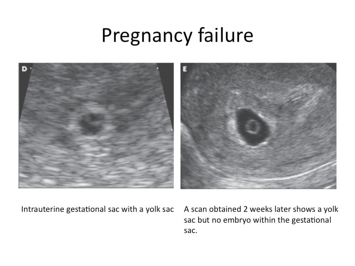
An imaging approach to early pregnancy failure

Early Pregnancy Ultrasound Assessment of Multiple Pregnancy | IntechOpen

Diagnostic Ultrasound in the First Trimester of Pregnancy | GLOWM

Ultrasound is a Critical Tool of Managing Miscarriage

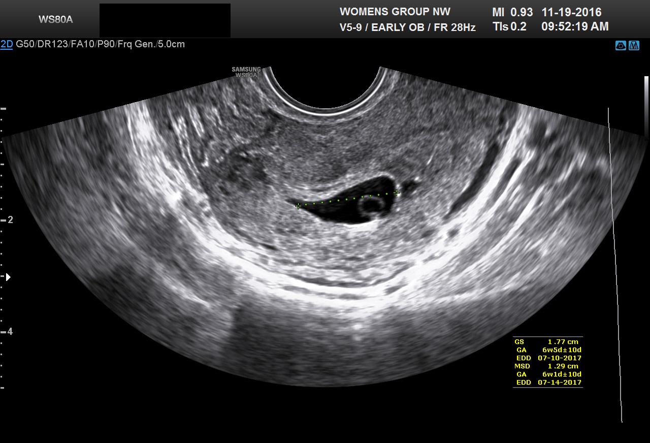
Ultrasound Video showing an empty Gestational sac in pregnancy of about 6  weeks 2 days. - YouTube
Ultrasound Video showing an empty Gestational sac in pregnancy of about 6 weeks 2 days. - YouTube

Abnormal Ultrasound in Early Pregnancy

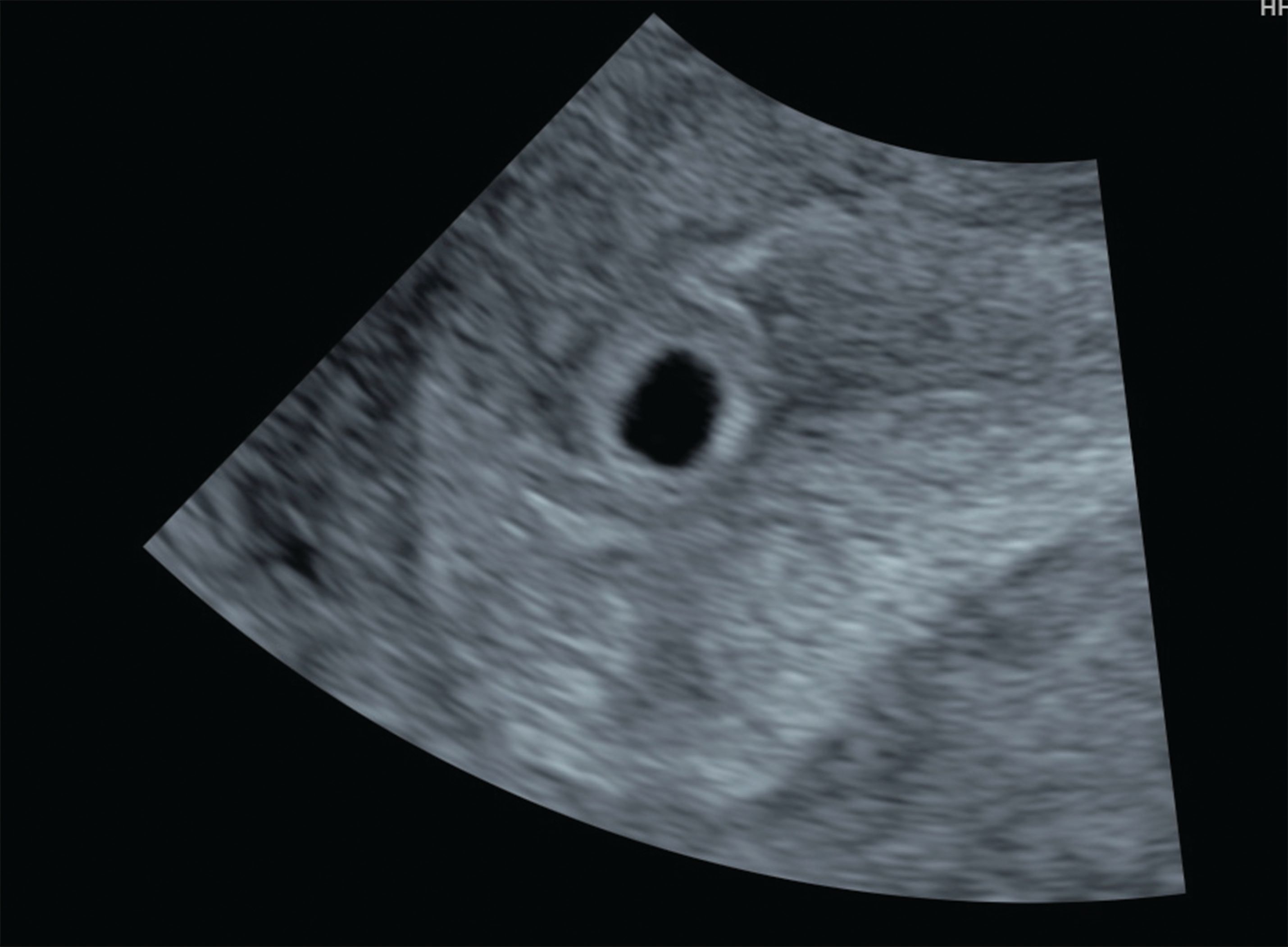
Gestational sacs from 3-4 mm of diameter can usually be seen. | Download  Scientific Diagram
Gestational sacs from 3-4 mm of diameter can usually be seen. | Download Scientific Diagram

Difference between gestational sac and yolk sac? | Difference Between

Table 4 from Size of Yolk Sac by Ultrasonography and Its Correlation with  Pregnancy Outcome | Semantic Scholar
Table 4 from Size of Yolk Sac by Ultrasonography and Its Correlation with Pregnancy Outcome | Semantic Scholar

Ultrasonography showing gestational sac of 4 weeks and 6 days. | Download  Scientific Diagram
Ultrasonography showing gestational sac of 4 weeks and 6 days. | Download Scientific Diagram

The Gestational Sac In Pregnancy | babyMed.com

Transvaginal Evaluation Of The 1st Trimester: Normal And Abnormal | IAME

Yolk sac | Radiology Reference Article | Radiopaedia.org

The Normal Gestational Sac

Diagnostic Ultrasound in the First Trimester of Pregnancy | GLOWM

Diagnostic Ultrasound in the First Trimester of Pregnancy | GLOWM

Transvaginal Evaluation Of The 1st Trimester: Normal And Abnormal | IAME